Hypdysplasia in dogs
**************************
Canine hip dysplasia is a very common degenerative joint disease seen in dogs.
There are many misconceptions surrounding it. There are many things that we
know about hip dysplasia in dogs, there are also many things we suspect about
this common cause of limping, and there are some things that we just do not
know about the disease. We will cover all of those here and hope to separate
out fact, theory, hypothesis, and opinion.
What is hip dysplasia?
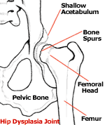
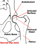
To understand what hip dysplasia really is we must have a basic understanding
of the joint that is being affected. The hip joint forms the attachment of the
hind leg to the body and is a ball and socket joint. The ball portion is the
head of the femur while the socket (acetabulum) is located on the pelvis. In a
normal joint, the ball rotates freely within the socket. To facilitate
movement, the bones are shaped to perfectly match each other, with the socket
surrounding the ball. To strengthen the joint, the two bones are held together
by a ligament. The ligament attaches the femoral head directly to the
acetabulum. Also, the joint capsule, which is a very strong band of connective
tissues, encircles the two bones adding further stability. The area where the
bones actually touch each other is called the articular surface. It is
perfectly smooth and cushioned with a layer of spongy cartilage. In the normal
dog, all of these factors work together to cause the joint to function
smoothly and with stability.
Hip dysplasia results from the abnormal development of the hip joint in the
young dog. It may or may not be bilateral, affecting both right and left
sides. It is brought about by the laxity of the muscles, connective tissue,
and ligaments that should support the joint. Most dysplastic dogs are born
with normal hips, but due to genetic and possibly other factors, the soft
tissues that surround the joint start to develop abnormally as the puppy
grows. The most important part of these changes is that the bones are not held
in place, but actually move apart. The joint capsule and the ligament between
the two bones stretch, adding further instability to the joint. As this
happens, the articular surfaces of the two bones lose contact with each other.
This separation of the two bones within a joint is called subluxation and
this, and this alone, causes all of the resulting problems we associate with
the disease.
What are the symptoms
of hip dysplasia?
Dogs of all ages are subject to the symptoms of hip dysplasia and the
resultant osteoarthritis. In severe cases, puppies as young as five months
will begin to show pain and discomfort during and after vigorous exercise. The
condition will worsen until even normal daily activities are painful. Without
intervention, these dogs may be unable to walk at all by a couple years of
age. In most cases, however, the symptoms do not begin to show until the
middle or later years in the dog’s life.
The symptoms are typical for those seen with other causes of osteoarthritis.
Dogs may walk or run with an altered gait, often resisting movements that
require full extension or flexion of the rear legs. Many times, they run with
a ‘bunny hopping’ gait. They will show stiffness and pain in the rear legs
after exercise or first thing in the morning. Most dogs will warm up out of
the muscle stiffness with movement and exercise. Some dogs will limp and many
will decrease their level of activity. As the condition progresses, the dogs
will lose muscle tone and may even need assistance in getting up. Many owners
attribute the changes to normal aging, but after treatment is initiated, they
are shocked to see much more normal and pain-free movement return.
Who gets hip
dysplasia?
Hip dysplasia can be found in dogs, cats, and humans, but for this article, we
are concentrating only on dogs. In dogs, it is primarily a disease of large
and giant breeds. The disease can occur in medium-sized breeds and rarely even
in small breeds. It is primarily a disease of purebreds although it can happen
in mixed breeds, particularly if it is a cross of two dogs that are prone to
developing the disease. German Shepherds, Labrador Retrievers, Rottweillers,
Great Danes, Golden Retrievers, and St. Bernards appear to have a higher
incidence, however, these are all very popular breeds and may be over
represented because of their popularity. On the other end Greyhounds and
Borzois have a very low incidence of the disease.
What are the risk
factors for the development of hip dysplasia?
Hip dysplasia is caused by looseness in the hip joint. The looseness creates
abnormal wear and erosion of the joint and as a result pain and arthritis
develops. The disease process is fairly straightforward; the controversy
starts when we try to determine what predisposes animals to contract the
disease. Almost all researchers agree that there is a genetic link involved.
If a parent has hip dysplasia, then the offspring are at greater risk for
developing hip dysplasia. Some researchers feel that genetics are the only
factor involved, where others feel that genetics contribute less than 25% to
the development of the disease. The truth probably lies in the middle. If
there are no carriers of hip dysplasia in a dog’s lineage, then it will not
contract the disease. If there are genetic carriers, then it may contract the
disease. We can greatly reduce the incidence of hip dysplasia through
selective breeding. We can also increase the incidence through selectively
breeding. We cannot, however, completely reproduce the disease through
selective breeding. In other words, if you breed two dysplastic dogs, the
offspring are much more likely to develop the disease, but will not all have
the same level of symptoms or even necessarily show any symptoms. The
offspring from these dogs will, however, be carriers and the disease may show
up in their offspring in later generations. This is why it can be difficult to
eradicate the disease from a breed or specific line.
Nutrition: Experimentally, we can increase the
severity of the disease in genetically susceptible animals in a number of
ways. One of them is through obesity. It stands to reason that carrying around
extra weight will exacerbate degeneration of the joint in a dog with a loose
hip. Overweight dogs are therefore at a much higher risk. Another factor that
may increase the incidence is rapid growth in a puppy during the ages from
three to ten months. Experimentally, the incidence has been increased in
genetically susceptible dogs when they are given free choice high protein,
high calorie diets. In a large study done in 1997, Labrador Retriever puppies
fed a high protein, high calorie diet free choice for three years had a much
higher incidence of hip dysplasia than their litter mates who were fed the
same high calorie, high protein diet, but in an amount that was 25% less than
that fed to the dysplastic group. As might be expected, however, the free
choice group was significantly heavier at maturity and averaged 22 pounds
heavier than the control group. Because obesity is also a risk factor, this
study may be difficult to interpret.
|
I have yet to see a
study that links an increased incidence of hip dysplasia in dogs fed a
normal diet of commercial puppy food versus a specialty diet formulated for
just large breed dogs. |
There have also been studies looking into protein and calcium levels and their
relationship to hip dysplasia. Both of these studies were able to increase the
level of hip dysplasia by feeding increased amounts of calcium and protein.
But once again, the studies of puppies fed greatly increased amounts over
normal recommended values and compared them to animals fed decreased amounts.
They failed to compare puppies fed a normal amount of food that had the
recommended amount of protein, fat, and calcium to those fed a diet with
slightly less protein, fat, and calcium (similar to those 'large breed puppy
foods' that are now flooding the market). I have yet to see a study that links
an increased incidence in hip dysplasia in dogs fed a normal diet of
commercial puppy food versus a specialty diet formulated just for large breed
puppies.
Exercise: Exercise may be another risk factor.
It appears that dogs that are genetically susceptible to the disease may have
an increased incidence of disease if they over-exercise at a young age. But at
the same time, we know that dogs with large and prominent leg muscle mass are
less likely to contract the disease than dogs with small muscle mass. So
exercising and maintaining good muscle mass may actually decrease the
incidence of the disease. Moderate exercise that strengthens the gluteal
muscles, such as running and swimming, is probably a good idea. Whereas,
activities that apply a lot of force to the joint are contraindicated. An
example would be a jumping activity such as playing Frisbee.
How is hip dysplasia
diagnosed?
Diagnosis of hip dysplasia in dogs that are showing clinical signs of
arthritis and pain is usually made through the combination of a physical exam
and radiographs (x-rays). If a dog is showing outward signs of arthritis,
there are usually easily recognized changes in the joint that can be seen on
radiographs. In addition, the veterinarian may even be able to feel looseness
in the joint or may be able to elicit pain through extension and flexion.
Regardless, the results are straightforward and usually not difficult to
interpret.
However, about half of the animals that come in for a determination on the
health of their hip joints are not showing physical signs, but are intended to
be used for breeding. The breeder wants to ensure that the animal is not at
great risk for transmitting the disease to his or her offspring. There are two
different testing methods that can be performed. The traditional and still
most common is OFA testing. The other newer technique is the PennHip method.
OFA: The method used by the Orthopedic
Foundation for Animals (OFA) has been the standard for many years.
The OFA was established in 1966, and has become the world’s largest all-breed
registry. The OFA maintains a database of hip evaluations for more than
475,000 dogs. Radiographs are taken by a local veterinarian under specific
guidelines and are then submitted to the OFA for evaluation of hip dysplasia
and certification of hip status. Since the accuracy of radiological diagnosis
of hip dysplasia using the OFA technique increases after 24 months of age, the
OFA requires that the dog be at least two years of age at the time the
radiographs are taken. They also recommend that the evaluation should not be
performed while the female is in heat. To get the correct presentation and
ensure that the muscles are relaxed, the OFA recommends that the dog be
anesthetized for the radiographs. OFA radiologists evaluate the hip joints for
congruity, subluxation, the condition of the acetabular margins and acetabular
notch, and the size, shape, and architecture of the femoral head and neck. The
radiographs are reviewed by three radiologists and a consensus score is
assigned based on the animal's hip conformation relative to other individuals
of the same breed and age. Using a seven point scoring system, hips are scored
as normal (excellent, good, fair), borderline dysplastic, or dysplastic (mild,
moderate, severe). Dogs with hips scored as borderline or dysplastic are not
eligible to receive OFA breeding numbers.
|
When dogs born in
1972 to 1980 were compared with dogs born in 1989 and 1990, 60% of the
breeds demonstrated a statistically significant decrease in hip dysplasia.
At the same time, 68% of breeds had a statistically significant increase in
the number of hips scored as excellent. |
The
OFA will also provide preliminary evaluations (performed by one OFA
radiologist) of dogs younger than 24 months of age to help breeders choose
breeding stock. Reliability of the preliminary evaluation is between 70 and
100% depending on the breed. Results published by the OFA suggest that the
incidence of hip dysplasia in certain breeds has decreased as a result of
selective breeding programs. When dogs born in 1972 to 1980 were compared with
dogs born in 1989 and 1990, 60% of the breeds demonstrated a statistically
significant decrease in hip dysplasia. At the same time, 68% of breeds had a
statistically significant increase in the number of hips scored as excellent.
This information may suggest progress is being made to decrease the frequency
of hip dysplasia, but it may simply be that only radiographs from dogs thought
to have normal hips are being submitted to the OFA, while those with dysplasia
are being screened out by referring veterinarians.
PennHIP: The diagnostic method used by the
University of Pennsylvania Hip Improvement Program (PennHIP)
uses distraction/compression radiographic views to more accurately identify
and quantify joint laxity. Radiographs of the hip joints are taken with the
dog under heavy sedation. Two views are obtained with the hind limbs in
neutral position to maximize joint laxity. Weights and an external device are
used to help push the head of the femur further into or away from the
acetabulum. The amount of femoral head displacement (joint laxity) is
quantified using a distraction index (DI). The DI ranges from 0 to 1 and is
calculated by measuring the distance the center of the femoral head moves
laterally from the center of the acetabulum and dividing it by the radius of
the femoral head. A DI of 0 indicates a very tight joint. A DI of 1 indicates
complete luxation with little or no coverage of the femoral head. A hip with a
distraction index of .6 is 60% luxated and is twice as lax as a hip with a DI
of .3. When the DI was compared to the OFA scores for 65 dogs, all dogs scored
as mildly, moderately, or severely dysplastic by the OFA method had a DI above
.3.
Hip joint laxity as measured by the DI is strongly correlated with the future
development of osteoarthritis. Hips with a low DI are less likely to develop
osteoarthritis. Hips with a DI below .3 rarely develop osteoarthritis visible
on radiographs. Although hips with a DI above .3 are considered "degenerative
joint disease susceptible," not all hips with a DI greater than .3 eventually
develop osteoarthritis. It is known that some hips with radiographically
apparent laxity do not develop osteoarthritis. A means of differentiating lax
hips that develop osteoarthritis from those that will not is important in
developing a prognosis and making treatment recommendations. In one study, the
DI obtained from dogs at four months of age was a good predictor of later
osteoarthritis, though the 6 and 12-month indices were more accurate.
To assure quality and repeatability among diagnostic centers using the PennHip
technique, veterinarians must take a special training course to become
certified. As this technique gains popularity more and more veterinarians are
becoming certified.
How is hip dysplasia
treated?
Surgical Treatment of
Hip Dysplasia:
There are several surgical procedures available depending on the age and the
severity of the joint degeneration.
Triple Pelvic Osteotomy (TPO): TPO is a
procedure used in young dogs usually less than 10 months of age that have
radiographs that show severe hip laxity but have not developed severe damage
to the joints. The procedure involves a surgical breaking of the pelvic bones
and a realignment of the femoral head and acetabulum restoring the coxofemoral
weight bearing surface area and correcting femoral head subluxation. This is a
major surgery and is very expensive but the surgery has been very successful
on animals that meet the requirements.
Total Hip Replacement: may be the best
surgical option for dogs that have degenerative joint disease as a result of
chronic hip dysplasia. Total hip replacement is a salvage procedure that can
produce a functionally normal joint, eliminate degenerative changes and
alleviate joint pain. The procedure involves the removal of the existing joint
and replacing it with a prosthesis. To be a candidate for this procedure the
animal must be skeletally mature and weigh at least 35 pounds. There is no
maximum size limit. If both hips need to be replaced there is a three-month
period of rest recommended between the surgeries. As with the TPO surgery this
is a very expensive procedure but has had some very good results.
Femoral Head and Neck Excision: Femoral head
and neck excision is a procedure in which the head of the femur is surgically
removed and a fibrous pseudo-joint forms. This procedure is considered a
salvage procedure and is used in cases where degenerative joint disease has
occurred and total hip replacement isn’t feasible. The resulting pseudo-joint
will be free from pain and allow the animal to increase its activity, however,
full range of motion and joint stability are decreased. For best results the
patient should weigh less than 50 pounds, however the procedure is often
performed on larger dogs.
Pectineal Myectomy: This is a somewhat
controversial treatment for patients with chronic hip dysplasia. The pectineus
is one of the muscles attaching the femur to the pelvis. By cutting and
removing this muscle, the tension on the joint and joint capsule are reduced.
This offers some pain relief for some patients but doesn’t slow the
progression of the disease. There are possible complications with this
procedure and with the introduction of the newer, better procedures this
surgery is rarely performed anymore.
Medical Treatment of
Hip Dysplasia:
|
Because hip
dysplasia is primarily an inherited condition, there are no products on the
market that prevent the development of hip dysplasia. |
Medical treatment has greatly improved in the last several years thanks to the
introduction and approval of several new drugs used to treat osteoarthritis.
Because hip dysplasia is primarily an inherited condition there are no
products on the market that prevent the development of hip dysplasia. I very
often get asked if a certain product will prevent hip dysplasia but I always
must answer "no". Through proper diet, exercise and supplemental glucosamine
you can decrease the progression of degenerative joint disease but the
looseness in the joint will not change significantly as a result of any
supplement.
Medical management is indicated for young dogs with a sudden onset of clinical
signs and for older dogs with chronic osteoarthritis. Because of the high cost
involved with many surgeries, medical management is many times the only
realistic option for many clients. Medical management is a multifaceted
treatment. For the best results, several of the following treatments must be
instituted. For most animals, I began with the first recommendations and work
my way down this list as needed to control the pain and degenerative joint
disease.
Weight Management: Weight management is the
first thing that must be addressed. All surgical and medical procedures will
work much better if the animal is not overweight. Considering that up to half
of the pets in the U.S. are overweight there is a fair chance that many of the
dogs with hip dysplasia are also overweight. Getting the dog down to it’s
recommended weight and keeping it there may be the most important thing an
owner can do for their pet. However this may be the hardest part of the
treatment, but it’s worth it. Very few dogs can drive to MacDonalds, work a
can opener, or open the refrigerator, so you the owner are controlling what
the dog eats. If you feed your dog less it will lose weight.
Exercise: Exercise is the next important
step. What we are trying to accomplish here is to restrict the amount of
exercising yet still maintain adequate movement to increase or maintain muscle
strength. Young active dogs are going to need to be restricted to walks on the
leash or short periods of swimming. Older dogs should also participate in
these activities to a lesser extent. Too little exercise can be more
detrimental than too much in some cases so make sure your dog is getting out
daily for some activity. Jumping in all forms is bad for dogs with hip
dysplasia. While watching a dog play Frisbee is very enjoyable and fun for the
dog, remember that it is very hard on a dog's hip joints. Remember, it is
important to exercise daily; only exercising on weekends, for instance
may cause more harm than good if the animal is sore for the rest of the week
and reluctant to move at all.
Glucosamine and Chondroitin:Glucosamine and
chondroitin are two of the drugs that have recently become widely used
in treating both animals and humans for osteoarthritis. These products have
been around for a while but due to the lack of scientific studies supporting
them and the medical profession's resistance to endorse a nutraceutical, they
had failed to gain popularity. But due to the overwhelming success in treating
patients with osteoarthritis these products have come to the forefront of
therapy and are becoming one of the most popular products for treating
arthritis today.
Glucosamine is the major sugar found in glycosaminoglycans and hyaluronate,
which are important building blocks in the synthesis and maintenance of
cartilage in the joint. Chondroitin enhances the synthesis of
glycosaminoglycans and inhibits damaging enzymes in the joint.
When a dog has hip dysplasia the joint wears abnormally and the protective
cartilage on the surface of the joint gets worn away and the resultant bone to
bone contact creates pain. Glucosamine and chondroitin give the
cartilage-forming cells (chondrocytes) the building blocks they need to
synthesize new cartilage and to repair the existing damaged cartilage. These
products are not painkillers; they work by actually healing the damage that
has been done. These products generally take at least six weeks to begin to
heal the cartilage and most animals need to be maintained on these products
the rest of their lives to prevent further cartilage breakdown. Because these
products are naturally-occurring compounds they are very safe and show very
few side effects. There are many different glucosamine/chondroitin products on
the market but they are not all created equal. We have seen the best results
and fewest side effects from products that are formulated especially for dogs
and that contain pure ingredients that are human grade in quality. Products
such as Dr. Foster and Smith's Joint Care and Gluco-C, or the veterinary-sold
product Cosequin are several that fit this category.
Buffered Aspirin: Buffered aspirin is an
excellent anti-inflammatory and painkiller in dogs (Do NOT give your cat
aspirin unless prescribed by your veterinarian). It can be used along with
glucosamine/chondroitin products and is safe for long term use. With all
aspirin products used in dogs there is a risk of intestinal upset or in rare
cases gastric ulceration. Because of these problems it is recommended that if
a dog develops signs of GI upset, the product be discontinued until a
veterinary exam can be performed. (By giving aspirin with a meal, you may be
able to reduce the possibility of side effects.) Using buffered aspirin
formulated just for dogs makes dosage and administration much easier. Regular
aspirin, Tylenol, and ibuprofen have many more potential side effects and are
not recommended without veterinary guidance.
Carprofen (Rimadyl): Rimadyl is a
non-steroidal anti-inflammatory developed for use in dogs with osteoarthritis.
Carprofen is a very strong and effective pain killer and anti-inflammatory
agent. It is a prescription product and because of potential side effects,
careful adherence to dosing quantity and frequency must be followed. Many
veterinarians throughout the country are requiring periodic bloodwork to be
done on animals that are on this product to monitor any developing liver
problems resulting from its use. This product is often used initially with
glucosamine therapy and then as the glucosamine product begins to work the
carprofen dose is reduced or eliminated.
Polysulfated Glycosaminoglycan (Adequan):
Adequan is a product that is administered as an injection. A series of shots
are given over weeks and very often have favorable results. The cost and the
inconvenience of weekly injections are a deterrent to some owners especially
since the oral glucosamine products are so effective. This product helps
prevent the breakdown of cartilage and may help with the synthesis of new
cartilage. The complete mechanism of action of this product is not completely
understood but appears to work on several different areas in cartilage
protection and synthesis.
Vitamin C: Vitamin C has received a lot of
press lately, primarily because of studies done in humans that have linked it
to preventing and controlling a variety of diseases. Much of the use in
animals has been extrapolated from human medicine. Humans are one of only a
handful of species that have a requirement for vitamin C. Dogs and cats
synthesize their own vitamin C so this is one area where we probably shouldn’t
be using human studies as guidelines for treating cats and dogs. We know that
vitamin C acts as an antioxidant and is an important nutrient in the synthesis
of collagen and cartilage. We also know that Vitamin C is water-soluble and it
is very difficult to create a toxicity. Vitamin C does lower the pH of urine
and some researchers question the possible long term side effects of
over-acidified urine. The benefits of vitamin C in preventing or treating hip
dysplasia and joint disease are purely speculative. Using reasonable doses of
Vitamin C doesn’t appear to be harmful and some day research may show that it
is beneficial in animals.
Corticosteroids: Corticosteroids have been
used for many years to treat the pain and inflammation associated with
osteoarthritis. Corticosteroids act as a potent anti-inflammatory but
unfortunately have many undesirable short and long term side effects. Because
of these side effects and the advent of newer, more specific drugs,
corticosteroids are generally only used in older animals where all other pain
control products have failed. Corticosteroids are a prescription product and
come in both a pill and injectable form.
How do we prevent hip
dysplasia?
|
When it comes to
preventing the formation of hip dysplasia there is only one thing that all
researchers agree on, and that is selective breeding is crucial. |
There are many different theories on how to prevent the progression of hip
dysplasia. As I discussed earlier, nutrition, exercise, and body weight may
all contribute to the severity of degenerative joint disease after the hip
dysplasia has developed. When it comes to preventing the formation of hip
dysplasia there is only one thing that all researchers agree on, and that is
selective breeding is crucial. There will be a lot of new information coming
forward in the future concerning other factors that contribute to hip
dysplasia, but for right now we have to stick to what we know for sure. We
know that through selectively breeding animals with good hips we can
significantly reduce the incidence of hip dysplasia. We also know that we can
increase the incidence of hip dysplasia if we choose to use dysplastic animals
for breeding. Breeding two animals with excellent hips does not guarantee that
all of the offspring will be free of hip dysplasia but there will be a much
lower incidence than if we breed two animals with fair or poor hips. If we
only bred animals with excellent hips it wouldn’t take long to make hip
dysplasia a rare occurrence. If owners insisted on only purchasing an animal
that had parents and grandparents with certified good or excellent hips, or if
breeders only bred these excellent animals then the majority of the problems
would be eliminated. For the best results, buyers should look at three or four
generations of dogs prior to theirs to insure that there aren’t carriers in
the bloodline. Following the newer recommendations for exercise and nutrition
may help but will never come close to controlling or eliminating the disease
if stricter requirements for certified hips aren’t instituted or demanded.
Summary
Hip Dysplasia is a widespread condition that primarily affects large and giant
breeds of dogs. There is a strong genetic link between parents that have hip
dysplasia and the incidence in their offspring. There are probably other
factors that contribute toward the severity of the disease.
Osteoarthritis is the result of degeneration of the joint due to hip
dysplasia. Surgical and medical treatments are targeted to prevent and treat
the resulting osteoarthritis. The best way to prevent hip dysplasia is through
selection of offspring whose parents and grandparents have been certified to
have excellent hip conformation.